
购物车0
产品总数:60942


| 中文名称 |
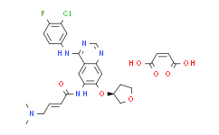
Afatinib dimaleate.
|
|---|---|
| 中文别名 |
双马来酸盐阿法替尼;双马来酸阿法替尼;阿法替尼AfatinibBIBW2992;阿法替尼二马来酸盐;阿法替尼双马来酸盐;阿法替尼双马来酸盐 仅供科研使用;马来酸阿法替尼;双马来酸阿法替尼(BIBW2992);2-(二甲基亚膦酰基)苯胺;顺丁烯二酸盐
|
| 英文名称 |
Afatinib dimaleate
|
| 英文别名 |
2-Butenamide,
N-[4-[(3-chloro-4-fluorophenyl)amino]-7-[[(3S)-tetrahydro-3-furanyl]oxy]-
6-quinazolinyl]-4-(dimethylamino)-, (2E)-, (2Z)-2-butenedioate (1:2);Afatinib dimaleate;BIBW2992 DiMaleate;(2E)-N-(4-[(3-chloro-4-fluorophenyl)amino]-7-{[(3S)-tetrahydrofuran-3-yl]oxy}quinazolin- 6-yl)-4-(dimethylamino)but-2-e...;(2E)-N-(4-[(3-chloro-4-fluorophenyl)amino]-7-{[(3S)-tetrahydrofuran-3-yl]oxy}quinazolin- 6-yl)-4-(dimethylamino)but-2-en;(2E)-N-(4-[(3-chloro-4-fluorophenyl)amino]-7-{[(3S)-tetrahydrofuran-3-yl]oxy}quinazolin- 6-yl)-4-(dimethylamino)but-2-enamide bis[hydrogen (2Z)-but-2-enedioate];(S,E)-N-(4-((3-Chloro-4-fluorophenyl)amino)-7-((tetrahydrofuran-3-yl)oxy)quinazolin-6-yl)-4-(dimethylamino)but-2-enamide maleate;(Z)-but-2-enedioic acid,(E)-N-[4-(3-chloro-4-fluoroanilino)-7-[(3S)-oxolan-3-yl]oxyquinazolin-6-yl]-4-(dimethylamino)but-2-enamide;2-Butenamide,
N-[4-[(3-chloro-4-fluorophenyl)amino]-7-[[(3S)-tetrahydro-3-furanyl]oxy]-
6-quinazolinyl]-4-(dimethylamino)-,;2-Butenamide,
N-[4-[(3-chloro-4-fluorophenyl)amino]-7-[[(3S)-tetrahydro-3-furanyl]oxy]-
6-quin...;2-Butenamide,N-[4-[(3-chloro-4-fluorophenyl)amino]-7-[[(3S)-tetrahydro-3-furanyl]oxy]-6-quinazolinyl]-4-(dimethylamino)-, (2E)-, (2Z)-2-butenedioate (1:2);Afatinib (dimaleate);BIBW2992 Afatinib dimaleate;BIBW2992-MA2;BIBW2992;FR-179643;Afatinib;Afatinib (diMaleate), BIBW2992;Afatinib dimaleate (USAN);Afatinib maleate;Afatinib maleate (JAN);BIBW 2992MA2;BIBW-2992;Gilotrif (TN);BIBW2992-MA2 (Afatinib dimaleate);But-2-enedioic acid;(E)-N-[4-(3-chloro-4-fluoroanilino)-7-[(3S)-oxolan-3-yl]oxyquinazolin-6-yl]-4-(d
|
| Cas No. |
850140-73-7
|
| 分子式 |
C32H33ClFN5O11
|
| 分子量 |
718.08
|
| 包装储存 |
Sealed and stored at 4℃,, away from moisture *In solvent : -80°C, 6 months; -20°C, 1 month (sealed storage, away from moisture) |
| 生物活性 |
Afatinib (BIBW 2992) dimaleate is an orally active, potent and irreversible dual specificity inhibitor of ErbB family (EGFR and HER2), with IC50 values of 0.5 nM, 0.4 nM, 10 nM and 14 nM for EGFR, EGFR, EGFR and HER2, respectively. Afatinib dimaleate can be used for the research of esophageal squamous cell carcinoma (ESCC), non-small cell lung cancer (NSCLC) and gastric cancer. |
||||||||||||||||||||||||||||||||||||||||
|---|---|---|---|---|---|---|---|---|---|---|---|---|---|---|---|---|---|---|---|---|---|---|---|---|---|---|---|---|---|---|---|---|---|---|---|---|---|---|---|---|---|
| 性状 |
Solid |
||||||||||||||||||||||||||||||||||||||||
| IC50 & Target[1][2] |
|
||||||||||||||||||||||||||||||||||||||||
| 体外研究(In Vitro) |
Afatinib dimaleate (100 nM) sufficiently prevents heregulin-stimulated HER3 phosphorylation. Medlife has not independently confirmed the accuracy of these methods. They are for reference only. Cell Proliferation Assay
Cell Viability Assay
Western Blot Analysis
Cell Cycle Analysis
Apoptosis Analysis
|
||||||||||||||||||||||||||||||||||||||||
| 体内研究(In Vivo) |
Afatinib dimaleate (0-20 mg/kg, Orally, daily for 25 days) shows dramatic tumor regression and downregulation of EGFR, HER2, HER3 and AKT phosphorylation. Medlife has not independently confirmed the accuracy of these methods. They are for reference only.
|
||||||||||||||||||||||||||||||||||||||||
| 运输条件 |
Room temperature or refrigerated transportation. |
||||||||||||||||||||||||||||||||||||||||
| 储存方式 |
Sealed and stored at 4℃,, away from moisture *In solvent : -80°C, 6 months; -20°C, 1 month (sealed storage, away from moisture) |
||||||||||||||||||||||||||||||||||||||||
| ClinicalTrial |
|
||||||||||||||||||||||||||||||||||||||||
| 参考文献 |
|
| 溶解度数据 |
体外研究:
H2O : 50 mg/mL (69.63 mM; Need ultrasonic) DMSO : ≥ 35 mg/mL (48.74 mM) * "≥" means soluble, but saturation unknown. 配制储备溶液
*
产品不同,其溶解度不同。建议根据产品选择合适的溶剂配制储备溶液;配成溶液后,建议分装保存,避免反复冻融造成的产品失效。 体内研究:
建议根据您的实验动物和给药方式选择适当的溶解方案。以下溶解方案都建议先按照 体外研究 方式配制澄清的储备液,再依次添加助溶剂:
——为保证实验结果的可靠性,澄清的储备液可以根据储存条件,适当保存;体内实验的工作液,建议您现用现配,当天使用;
以下溶剂前显示的百
*
|
|---|
1:一般建议:溶解度为Medlife测试数据,可能与文献描述存在差异。这是由于生产工艺和批次不同产生的正常现象。为了使其更好的溶解,请用37℃加热试管并在超声波水浴中震动片刻。不同批次产品溶解度各有差异,仅做参考,具体以实验方案为准。
2:储存条件:粉末-20°C一般情况可以保存3年,溶于溶剂-80°C一般情况可以保存1年。不同产品及不同批次产品可能存在差异,请细致阅读产品信息,并辅助参考相关文献描述。
Copyright © 2025 陌孚医药 All rights reserved 未经授权禁止拷贝本站所有资料,如有违反,将追究法律责任。
 沪ICP备2023012080号 |
 ![]() 沪公网安备31011402010657号
沪公网安备31011402010657号
 扫码关注公众号
 产品使用指南
