


| 中文名称 |
甲磺酸去铁胺.
|
|---|---|
| 中文别名 |
甲磺酸去铁胺;甲磺酸去铁敏;甲磺酸除铁灵;甲磺酸去铁胺标准品;甲磺酸去铁胺标准品(JP);去铁铵;去铁铵 USP标准品;去铁胺甲磺酸盐 EP标准品;去铁胺甲磺酸酯;十一酸甘油三酯;云铁胺甲磺酸酯;DEFEROX胺甲磺酸盐;去铁胺甲磺酸盐;去铁铵/去铁敏
|
| 英文名称 |
Deferoxamine mesylate
|
| 英文别名 |
Deferoxamine mesylate;Deferoxamine mesylate salt;Deferoxamine (mesylate);EHNA;Deferoxamine Methanesulfonate;Desferrioxamine B mesylate;DFOM;DFOM,Deferoxamine methanesulfonate salt,Desferrioxamine mesylate salt;Deferoxamine methanesulfonate salt;Desferrioxamine mesylate salt;desferal;Prestwick_988;desferalmesylate;deferoxaminemesilate;deferoxaminebmesylate;DEFEROXAMINE MESYLATE;monomethanesulfonate(sa;desferrioxaminebmesylate;desferalmethanesulfonate
|
| Cas No. |
138-14-7
|
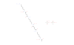
| 分子式 |
C27H56N6O14S2
|
| 分子量 |
752.90
|
| 包装储存 |
4°C, sealed storage, away from moisture *In solvent : -80°C, 6 months; -20°C, 1 month (sealed storage, away from moisture) |
| 生物活性 |
Deferoxamine mesylate (Deferoxamine B mesylate) is an iron chelator (binds to Fe(III) and many other metal cations), is widely used to reduce iron accumulation and deposition in tissues. Deferoxamine mesylate upregulates HIF-1α levels with good antioxidant activity. Deferoxamine mesylate also shows anti-proliferative activity, can induce apoptosis and autophagy in cancer cells. Deferoxamine mesylate can be used in studies of diabetes, neurodegenerative diseases as well as anti-cancer and anti-COVID-19. |
||||||||||||||||||||||||||||||||||||||||||||||||
|---|---|---|---|---|---|---|---|---|---|---|---|---|---|---|---|---|---|---|---|---|---|---|---|---|---|---|---|---|---|---|---|---|---|---|---|---|---|---|---|---|---|---|---|---|---|---|---|---|---|
| 性状 |
Solid |
||||||||||||||||||||||||||||||||||||||||||||||||
| 体外研究(In Vitro) |
Deferoxamine mesylate (1 mM; 16 h or 4 weeks) improves HIF-1α function under hypoxic and hyperglycemic conditions and decreases ROS in MEFs cells. Medlife has not independently confirmed the accuracy of these methods. They are for reference only. Western Blot Analysis
Western Blot Analysis
Cell Proliferation Assay
Apoptosis Analysis
Western Blot Analysis
Cell Autophagy Assay
|
||||||||||||||||||||||||||||||||||||||||||||||||
| 体内研究(In Vivo) |
Deferoxamine mesylate (560.68 mg/per; drip-on; once daily for 21 days) enhances wound healing and increases neovascularization in aged or diabetic mice. Medlife has not independently confirmed the accuracy of these methods. They are for reference only.
|
||||||||||||||||||||||||||||||||||||||||||||||||
| 运输条件 |
Room temperature or refrigerated transportation. |
||||||||||||||||||||||||||||||||||||||||||||||||
| 储存方式 |
4°C, sealed storage, away from moisture *In solvent : -80°C, 6 months; -20°C, 1 month (sealed storage, away from moisture) |
||||||||||||||||||||||||||||||||||||||||||||||||
| ClinicalTrial |
|
||||||||||||||||||||||||||||||||||||||||||||||||
| 参考文献 |
|
| 溶解度数据 |
体外研究:
H2O : 250 mg/mL (380.64 mM; Need ultrasonic) 配制储备溶液
*
产品不同,其溶解度不同。建议根据产品选择合适的溶剂配制储备溶液;配成溶液后,建议分装保存,避免反复冻融造成的产品失效。 体内研究:
建议根据您的实验动物和给药方式选择适当的溶解方案。以下溶解方案都建议先按照 体外研究 方式配制澄清的储备液,再依次添加助溶剂:
——为保证实验结果的可靠性,澄清的储备液可以根据储存条件,适当保存;体内实验的工作液,建议您现用现配,当天使用;
以下溶剂前显示的百
*
|
|---|
1:一般建议:溶解度为Medlife测试数据,可能与文献描述存在差异。这是由于生产工艺和批次不同产生的正常现象。为了使其更好的溶解,请用37℃加热试管并在超声波水浴中震动片刻。不同批次产品溶解度各有差异,仅做参考,具体以实验方案为准。
2:储存条件:粉末-20°C一般情况可以保存3年,溶于溶剂-80°C一般情况可以保存1年。不同产品及不同批次产品可能存在差异,请细致阅读产品信息,并辅助参考相关文献描述。
Copyright © 2025 陌孚医药 All rights reserved 未经授权禁止拷贝本站所有资料,如有违反,将追究法律责任。
 沪ICP备2023012080号 |
 ![]() 沪公网安备31011402010657号
沪公网安备31011402010657号
 扫码关注公众号
 产品使用指南
