
购物车0
产品总数:61739


| 中文名称 |
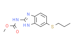
阿苯达唑.
|
||||||||||||
|---|---|---|---|---|---|---|---|---|---|---|---|---|---|
| 中文别名 |
丙硫咪唑;阿苯达唑;阿苯唑;扑尔虫;肠虫清;抗蠕敏;丙硫多菌灵悬浮剂;[5-(丙硫基)-1H-苯并咪唑-2-基]氨基甲酸甲酯;[5-(丙基硫代)-1H-苯并咪唑-2-基]氨基甲酸甲酯;5-丙硫基-苯并咪唑-2-氨基甲酸甲酯;丙硫苯咪唑;阿苯哒唑;阿苯哒唑 标准品;阿苯哒唑 杂质;阿苯达唑 EP标准品;阿苯达唑 USP标准品;阿苯达唑 标准品;阿苯达唑 ;丙硫达唑;阿苯达唑药典标准;氨基甲酸甲酯;丙硫咪唑 杂质对照品;丙硫咪唑原药;[5-(丙基硫代)苯并咪唑-2-基]氨基甲酸甲酯;5-(丙硫基)-2-苯并咪唑氨基甲酸甲酯;甲基[5-(丙基硫代)苯并咪唑-2-基]氨基甲酸酯;抗寄生虫;阿苯达唑杂质
|
||||||||||||
| 英文名称 |
Albendazole; SKF-62979
|
||||||||||||
| 英文别名 |
albendazole;Albendazole suspension;[5-(PROPYLTHIO)-1H-BENZIMIDAZOL-2-YL]CARBAMIC ACID, METHYL ESTER;[5-(PROPYLTHIO)BENZIMIDAZOL-2-YL]CARBAMIC ACID METHYL ESTER;(5-PROPYLSULFANYL-1H-BENZOIMIDAZOL-2-YL)-CARBAMIC ACID METHYL ESTER;ALBAZINE;ALBEN;ALBENZA;AKOS NCG1-0064;5-propylthio-2-carbomethoxyaminobenzimidazole;Atasol;Bilutac;Bruzol;methyl [5-(propylsulfanyl)-1H-1,3-benzimidazol-2-yl]carbamate;Methyl [5-(Propylthio)BenziMidazol-2-yl]CarbaMate;methyl 5-(propylthio)benzimidazol-2-ylcarbamate;methyl N-[5-(propylthio)-1H-benzimidazol-2-yl]carbamate;methyl-5-propylthio-2-benzimidazole carbamate;Monil;ZEBEN;ZENTEL;Methyl 5-propylthio-2-benzimidazolecarbamate;Eskazole;Valbazen;Proftril;Albendazolum;Albendazol;Zental;SK&F 62979;SKF 62979;Methyl 5-(propylthio)-2-benzimidazolecarbamate;Albendazolum [INN-Latin];Albendazol [INN-Spanish];5-(Propylthio)-2-carbomethoxyaminobenzimidazole;methyl N-[6-(propylsulfanyl)-1H-1,3-benzodiazol-2-yl]carbamate;(5-(Propylthio)-1H-benzimidazol-2-yl)carbamic acid methyl ester;O-Methyl N-(5-(propylthi
|
||||||||||||
| Cas No. |
54965-21-8
|
||||||||||||
| 分子式 |
C12H15N3O2S
|
||||||||||||
| 分子量 |
265.33
|
||||||||||||
| 包装储存 |
|
||||||||||||
| 详情描述 |
治疗寄生虫侵染,Albendazole属于苯并咪唑类化合物,可抗蠕虫感染。 |
| 生物活性 |
Albendazole (SKF-62979) is an orally active and broad-spectrum parasiticide with high effectiveness and low host toxicity, is used for the research of gastrointestinal parasites in humans and animals. Albendazole induces apoptosis and autophagy in cancer cells. Albendazole also inhibits tubulin polymerization and HIF-1α, VEGF expression, has antioxidant activity, and inhibits the glycolytic process in cancer cells. |
||||||||||||||||||||||||||||||||||||||||||||||||
|---|---|---|---|---|---|---|---|---|---|---|---|---|---|---|---|---|---|---|---|---|---|---|---|---|---|---|---|---|---|---|---|---|---|---|---|---|---|---|---|---|---|---|---|---|---|---|---|---|---|
| 性状 |
Solid |
||||||||||||||||||||||||||||||||||||||||||||||||
| IC50 & Target[1][2] |
parasites, tubulin. |
||||||||||||||||||||||||||||||||||||||||||||||||
| 体外研究(In Vitro) |
Albendazole (100, 500, 1000 nM; 1, 3, or 5 days) inhibits cell proliferation in a dose-dependent manner. Medlife has not independently confirmed the accuracy of these methods. They are for reference only. Cell Proliferation Assay
Cell Cycle Analysis
Apoptosis Analysis
Cell Autophagy Assay
Western Blot Analysis
Western Blot Analysis
|
||||||||||||||||||||||||||||||||||||||||||||||||
| 体内研究(In Vivo) |
Albendazole (10 mg/kg; i.g.; once a day for 30 days) reduces Echinococcus granulosus cyst weights in mice. Medlife has not independently confirmed the accuracy of these methods. They are for reference only.
|
||||||||||||||||||||||||||||||||||||||||||||||||
| 运输条件 |
Room temperature or refrigerated transportation. |
||||||||||||||||||||||||||||||||||||||||||||||||
| 储存方式 |
|
||||||||||||||||||||||||||||||||||||||||||||||||
| ClinicalTrial |
|
||||||||||||||||||||||||||||||||||||||||||||||||
| 参考文献 |
|
| 溶解度数据 |
体外研究:
DMSO : 20 mg/mL (75.38 mM; Need ultrasonic) H2O : 0.67 mg/mL (2.53 mM; Need ultrasonic) 配制储备溶液
*
产品不同,其溶解度不同。建议根据产品选择合适的溶剂配制储备溶液;配成溶液后,建议分装保存,避免反复冻融造成的产品失效。 体内研究:
建议根据您的实验动物和给药方式选择适当的溶解方案。以下溶解方案都建议先按照 体外研究 方式配制澄清的储备液,再依次添加助溶剂:
——为保证实验结果的可靠性,澄清的储备液可以根据储存条件,适当保存;体内实验的工作液,建议您现用现配,当天使用;
以下溶剂前显示的百
*
|
|---|
[1]. Pourgholami MH, et al. 体外研究 and in vivo suppression of growth of hepatocellular carcinoma cells by albendazole. Cancer Lett. 2001 Apr 10;165(1):43-9. [Information]
[2]. Jung YY, et al. Regulation of apoptosis and autophagy by albendazole in human colon adenocarcinoma cells. Biochimie. 2022 Jul;198:155-166. [Information]
[3]. Zhou F, et al. Albendazole inhibits HIF-1α-dependent glycolysis and VEGF expression in non-small cell lung cancer cells. Mol Cell Biochem. 2017 Apr;428(1-2):171-178. [Information]
[4]. Vural G, et al. Efficacy of novel albendazole salt formulations against secondary cystic echinococcosis in experimentally infected mice. Parasitology. 2020 Nov;147(13):1425-1432. [Information]
[5]. Li L, et al. Determination of albendazole and metabolites in silkworm Bombyx mori hemolymph by ultrafast liquid chromatography tandem triple quadrupole mass spectrometry. PLoS One. 2014 Sep 25;9(9):e105637. [Information]
1:一般建议:溶解度为Medlife测试数据,可能与文献描述存在差异。这是由于生产工艺和批次不同产生的正常现象。为了使其更好的溶解,请用37℃加热试管并在超声波水浴中震动片刻。不同批次产品溶解度各有差异,仅做参考,具体以实验方案为准。
2:储存条件:粉末-20°C一般情况可以保存3年,溶于溶剂-80°C一般情况可以保存1年。不同产品及不同批次产品可能存在差异,请细致阅读产品信息,并辅助参考相关文献描述。
Copyright © 2025 陌孚医药 All rights reserved 未经授权禁止拷贝本站所有资料,如有违反,将追究法律责任。
 沪ICP备2023012080号 |
 ![]() 沪公网安备31011402010657号
沪公网安备31011402010657号
 扫码关注公众号
 产品使用指南
