
购物车0
产品总数:60953


| 中文名称 |
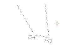
DiD perchlorate|细胞膜红色荧光探针
|
|---|---|
| 中文别名 |
1,1-双十八烷基-3,3,3,3-四甲基吲哚二碳菁;1,1'-双十八烷基-3,3,3',3'-四甲基吲哚二碳菁高氯酸盐;1,1`-双十八烷基-3,3,3`,3`-四甲基吲哚二碳花菁高氯酸盐;1,1'-双十八烷基-3,3,3',3'-四甲基吲哚二碳花菁高氯酸盐
|
| 英文名称 |
1,1'-DIOCTADECYL-3,3,3',3'-TETRAMETHYLINDODICARBOCYANINE PERCHLORATE
|
| 英文别名 |
1,1'-DIOCTADECYL-3,3,3',3'-TETRAMETHYLINDODICARBOCYANINE PERCHLORATE;DiD perchlorate;DiD( DiIC18(5));DiIC18(5) [DiD] [1,1'-Dioctadecyl-3,3,3',3'-tetramethylindodicarbocyanine perchlorate];DiIC18(5);1,1'-dioctadecyl-3,3,3',3'-tetrame.indo-dicarbocyanine clo4;1,1'-Dioctadecyl-3,3,3',3'-tetramethylindodicarbovyanineperchlorate;DID;'DID' OIL;DiD oil
DiIC18(5) oil;DIIC18(5) OIL;DILC18(5);'DID';D 307;dilC18(5) dye;Lipophilic Dye Did
|
| Cas No. |
127274-91-3
|
| 分子式 |
C61H99N2+.O4Cl-
|
| 分子量 |
959.90
|
| 包装储存 |
4°C, protect from light, stored under nitrogen 该产品在溶液状态不稳定,建议您现用现配,即刻使用。
|
| 产品详情 |
DiD 是一种长链碳菁染料。碳菁染料广泛用作亲脂性示踪剂,用于标记细胞、细胞器、脂质体、病毒和脂蛋白。
|
|---|---|
| 生物活性 |
DiD is a long-chain carbocyanine dye. Carbocyanine dyes are widely used as Di to label cells, organelles, liposomes, viruses and lipoproteins.
|
| 性状 |
Solid
|
| 体外研究(In Vitro) |
制备 Di 染色溶液1.1 制备 DMF、DMSO 或乙醇储备溶液:储备溶液应在 1-5 mM 的二甲基甲酰胺 (DMF) 、二甲基亚砜 (DMSO 或乙醇 DMSO 中制备。DMF 比乙醇更适合作为 Di 的溶剂。储备溶液应及时使用。任何未使用的溶液可作分装储存在 -20℃。避免反复冻融,溶液可储存 6 个月。1.2 制备工作溶液:将储备溶液稀释到合适的缓冲液中,如无血清培养基、HBS 或 PBS,以制备 1-5 μM工作溶液,现配现用。注:应根据经验确定不同实验条件下工作溶液的最终浓度。2. 悬浮细胞2.1 悬浮细胞:离心收集细胞,加入 PBS 洗涤两次,每次 5 分钟。细胞密度在 1×10/mL2.2 加入 1 mL Di 工作液,室温孵育 5-30 分钟。2.3 400 g,4℃ 离心 3-4 分钟,弃去上清。2.4 加入 PBS 洗涤细胞两次,每次 5 分钟。2.5 用 1 mL 无血清培养基或 PBS 重悬细胞后,使用荧光显微镜或流式细胞仪进行观察。3. 贴壁细胞3.1 将贴壁细胞培养于无菌盖玻片上。3.2 从培养基中移出盖玻片,吸除多余培养基。3.3 加入 100 μL 染料工作液,轻轻晃动使其完全覆盖细胞,孵育 5-30 分钟。3.4 吸去染料工作液,用培养基洗 2-3 次,每次 5 分钟,使用荧光显微镜或流式细胞仪进行观察。 has not independently confirmed the accuracy of these methods. They are for reference only.
|
| 体内研究(In Vivo) |
Two weeks after injection of stained cells, a single, bright, DiD perchlorate (DiD)-positive cells located between 15 to 40 μm from the endosteum is found. Results reveal a progressive appearance of cell clusters of decreased dye intensity, consistent with the partitioning of DiD perchlorate label on cell division. has not independently confirmed the accuracy of these methods. They are for reference only.
|
| 运输条件 |
Room temperature in continental US; may vary elsewhere.
|
| 储存方式 |
4°C, protect from light, stored under nitrogen 该产品在溶液状态不稳定,建议您现用现配,即刻使用。
|
| Emission(Em) |
Em 620-750 nm Red
|
| Excitation(Ex) |
Ex 620-750 nm Red
|
| 参考文献 |
[1]. Gan WB, et al. Multicolor "DiOlistic" labeling of the nervous system using lipophilic dye combinations. Neuron. 2000 Aug;27(2):219-25.[2]. Kenji Yumoto, et al. A novel method for monitoring tumor dormancy using fluorescent dye DiD. Cytometry A. 2014 Jun; 85(6): 548–555.[3]. Meng Li, et al. In Vivo Tracking of
|
| 溶解度数据 |
In Vitro: DMSO : 25 mg/mL (26.04 mM; ultrasonic and warming and heat to 60°C)配制储备液
|
|---|
1:一般建议:溶解度为Medlife测试数据,可能与文献描述存在差异。这是由于生产工艺和批次不同产生的正常现象。为了使其更好的溶解,请用37℃加热试管并在超声波水浴中震动片刻。不同批次产品溶解度各有差异,仅做参考,具体以实验方案为准。
2:储存条件:粉末-20°C一般情况可以保存3年,溶于溶剂-80°C一般情况可以保存1年。不同产品及不同批次产品可能存在差异,请细致阅读产品信息,并辅助参考相关文献描述。
Copyright © 2025 陌孚医药 All rights reserved 未经授权禁止拷贝本站所有资料,如有违反,将追究法律责任。
 沪ICP备2023012080号 |
 ![]() 沪公网安备31011402010657号
沪公网安备31011402010657号
 扫码关注公众号
 产品使用指南
